Augmented Reality in the Cath Lab: How Dr. Rahul Gupta Uses 3D Heart Models for Precision Surgery
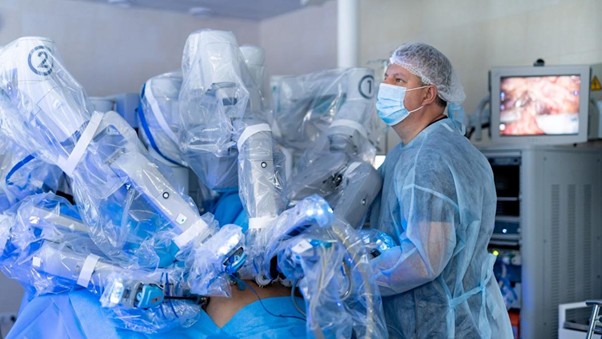
Imagine a surgeon who doesn’t just look at a monitor, but actually steps inside a holographic map of your heart before the procedure even begins. In 2026, the Cardiac Catheterization Lab has transformed from a room of flat, two-dimensional X-ray screens into a futuristic theater where complex surgeries are rehearsed and executed in a three-dimensional digital space. This isn’t science fiction—it is the new gold standard of precision heart care.
For decades, interventional cardiology relied on a fundamental challenge: the heart is a complex, three-dimensional moving object, but the tools used to see it—traditional angiography—provided only flat, 2D shadows. Surgeons had to mentally “reconstruct” the 3D depth of a patient’s arteries or valves based on these shadows. While expertise made this possible, the margin for error was always a factor.
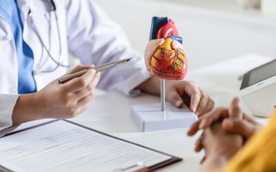
Today, the integration of Augmented Reality (AR) and 3D modeling has officially bridged that gap. By merging a patient’s specific CT or MRI data into a holographic “digital twin,” surgeons like Dr. Rahul Gupta can visualize the unique architecture of a heart with sub-millimeter precision. This shift is revolutionizing outcomes for patients in Navi Mumbai, Pune, and across the globe.
The Evolution of Visualization: From Shadows to Holograms
To understand the impact of AR in the Cath Lab, we must look at how far we have come. In a traditional “Cath Lab” (Catheterization Laboratory), cardiologists use catheters—thin, flexible tubes—to treat blockages or structural issues.
Historically, the “eyes” of the cardiologist were X-ray images. While life-saving, X-rays involve radiation and the use of contrast dye, and they only show the “vessel” as a dark silhouette against a white background.
The AR Difference
Augmented Reality allows the clinical team to overlay a 3D digital model of the patient’s specific anatomy directly onto the patient during the procedure. Using specialized headsets, the cardiologist sees a floating, high-definition hologram of the heart. They can rotate it, zoom in on a specific blockage, and see exactly where a stent or valve needs to be placed.
This “X-ray vision” eliminates the guesswork. It allows for a level of spatial awareness that was previously impossible, making procedures shorter, safer, and far more accurate.
Precision Surgery: The “Digital Twin” Strategy
One of the most profound applications of 3D modeling in 2026 is the creation of a “Digital Twin.” Before the patient even enters the Cath Lab at Cardium, their diagnostic data is used to create a virtual replica of their heart.
Dr. Rahul Gupta and the team can use this model to:
- Rehearse Complex Cases: For a patient with a “twisty” or heavily calcified artery, the team can virtually test different sizes of stents or different approach angles to see which one works best.
- Optimize Stent Placement: AI algorithms within the AR software can predict how a stent will expand in a specific area, ensuring that it fits perfectly against the vessel wall to prevent future blood clots.
- Reduce Radiation and Contrast: Because the surgeon has a “holographic map” to follow, they need fewer X-ray snapshots and less contrast dye to find their way, which is significantly healthier for the patient’s kidneys and overall system.
TAVI and Structural Heart Repair: A Natural Fit for AR
Nowhere is the precision of 3D modeling more critical than in structural heart repairs, such as Transcatheter Aortic Valve Implantation (TAVI).
In a TAVI procedure, a new valve is delivered through a catheter to replace a narrowed aortic valve. The sizing must be perfect. If the valve is a fraction of a millimeter too small, it might leak; if it’s too large, it could damage the heart tissue.
By using AR-guided 3D modeling, the team can visualize the exact “landing zone” for the new valve. They can see the calcification patterns in 3D and ensure the new valve is deployed with absolute exactness. This technology has turned TAVI into a highly predictable, “scar-less” success story for seniors who were previously considered too high-risk for surgery.
The Clinical Consensus: Why Depth Matters
Recent research published in leading academic forums, such as AHA Journals and Springer, confirms that 3D visualization significantly reduces “procedural time” and “complication rates.” In the world of cardiology, time is muscle. Every minute saved in the Cath Lab is a minute where the heart is under less stress.
Furthermore, the 2026 medical consensus highlights that precision surgery isn’t just about the procedure itself—it’s about the holistic data surrounding the patient. This includes understanding the patient’s metabolic health and nutritional markers (as explored in recent MDPI studies). When a high-tech AR procedure is combined with a data-driven lifestyle plan, the longevity of the repair is significantly enhanced.
At Cardium Heart Care the integration of technology is always balanced with authoritative, human expertise. AR is a powerful tool, but it is the experience of the cardiologist that interprets the data and makes the life-saving decisions.
FAQ: Understanding AR in Heart Surgery
Q: Does the AR headset distract the doctor during surgery?
A: Not at all. The technology is designed to be “heads-up.” Instead of having to look away from the patient at a distant monitor, the critical data is displayed right in the doctor’s line of sight, allowing them to keep their focus entirely on the patient.
Q: Is this technology only for “experimental” cases?
A: No. In 2026, 3D modeling and AR-assisted visualization are becoming the standard for complex stenting and structural repairs. It is used whenever the anatomy of the patient requires a higher level of precision than traditional X-rays can provide.
Q: Will I be able to see my 3D heart model?
A: Yes. One of the best uses of this technology is patient education. Dr. Rahul Gupta can show you your “Digital Twin,” explaining exactly where the blockage is and how the procedure will fix it. This transparency significantly reduces patient anxiety and helps in making informed decisions.
The Future of Heart Care: Predictive and Precise
The Cath Lab of 2026 is no longer just a place where we fix problems; it is a place where we use data to predict and prevent them. The “Virtual Safety Net” provided by AR and AI-guided imaging ensures that every patient receives a “custom-fit” solution.
Whether you are seeking a stress test , to evaluate your current risk or are preparing for a complex intervention, the goal of modern cardiology is to ensure you spend less time in the hospital and more time enjoying your life.
By merging the cutting edge of Augmented Reality with the authoritative expertise of a master clinician, we are removing the “shadows” from heart care. We are seeing the heart for what it truly is: a complex, vital organ that deserves the most precise, data-backed care science can offer.
Take the first step toward a clearer, more precise cardiovascular future. Explore how the latest technology can safeguard your heart at Cardium.
Contact Information (NAP)
Dr. Rahul Gupta Cardium Heart Care
Address: Shop No. 1 & 2, Intercity CHS, Swami Samarth Marg, behind Fire Brigade, Juhu Nagar, Sector-16, Vashi, Navi Mumbai, Maharashtra 400703
Phone no: 09869325666
Recent Posts
-
 Augmented Reality in the Cath Lab: How Dr. Rahul Gupta Uses 3D Heart Models for Precision Surgery
-
 The “Virtual Safety Net”: How Cardium’s AI-Powered Remote Monitoring Prevents Hospital Visits
-
 Stem Cell Therapy 2026: Can We Actually Regenerate a Damaged Heart Muscle?
-
 Pacemakers in 2026: Why the Latest Leadless Devices are Practically Invisible Under the Skin
-
 The 10-Minute TMT: What Your Stress Test Result Actually Says About Your 10-Year Heart Risk